
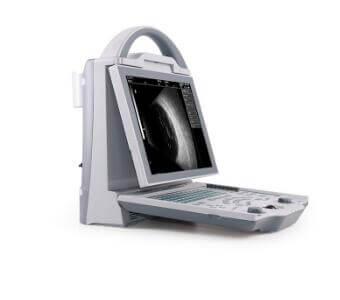
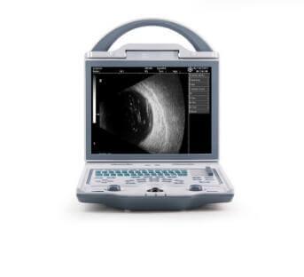
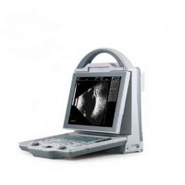
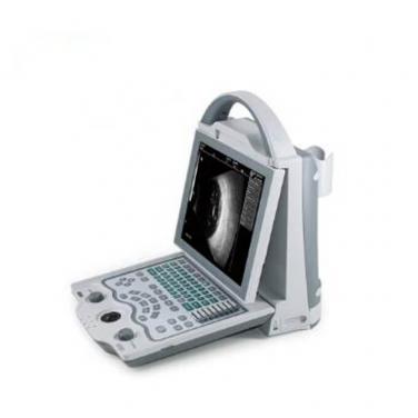
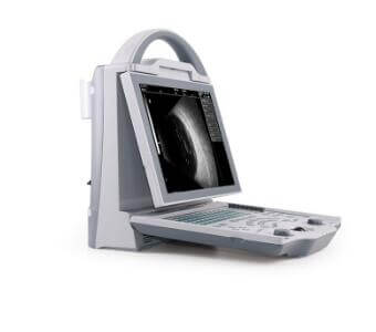
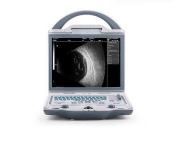
ODU 5 optical instruments ultrasonic scanner


 ODU5 technic parameter.pdf
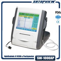
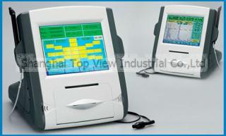
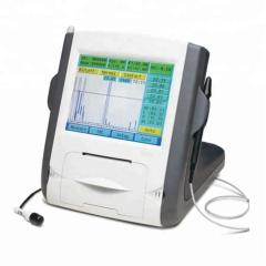
Ophthalmic A/B scan ODU5
General Parameter:A scan parameter:
| Resolution | 0.01mm |
| Gain adjustable | 0-120dB |
| Measurement range | 12-43mm (1640m/s) |
| Accuracy | ±0.02mm |
| Eye mode | normal / aphakia / dense cataract / PMMA, Acrylic & Silicon for Pseudo Phakic eyes |
| Measuring Mode | Contact and Immersion, automatic and manual |
| Measurement methods | A single-point measurement (under A mode); five-point measurement (under A mode) |
| Calculation |
std. deviation, average, automatical measuring 8 groups and average, accompany waveform, results correctable |
| IOL Formula | SRK-II, SRK-T, BINKHORST, HOLLADAY, HOFFER-Q, HAIGIS |
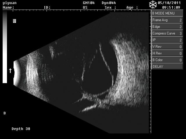
B scan parameter:
| Scan mode | High accuracy stepping motor drive sector scan |
| Scan Angle | 53° |
| Display Depth | 0~56mm |
| Gray | 256 |
| Depth seletion | 8 |
| Frame correlation | 4 |
| Edge enhancement | 4 |
| Compression Curve | 4 |
| Post-processing | 8 |
| image storage | 100 frames permanent image storage |
| General Measurement |
multi-electronic-rule to measure distance, circumference, area (trace method, ellipse method), volume (ellipse method), angle, histogram, profile, stenosis and curvature etc |
| B Scan |
| 10MHz probe, 10 frames/sec |
| Scanning Angle: 53° |
| Gain: 0~120dB adjustable, 8 points manually adjustable TGC |
| Distance, circumference, area, volume and angle measurement |
| Pesodu-color: 8 kinds |
| Cineloop: 128 frames, measurement available |
| Post processing: 4 groups of curves |
| A Scan |
| 10MHz with Fixation Red Light |
| Resolution: 0.01mm |
| Accuracy: t0.1mm |
| Measuring Range: 15~35mm |
| Measuring parameters: ACD, Lens, Vitreous body, AL |
| Measuring method: Contact and Immersion |
| Modes: Normal, Aphakic, Dense cataract, PMMA, Acrylic and Silicon |
| IOL formulas: SRK-T, SRK1\ BINKHORST, HOLLADAY, HOFFER-Q, HAIGIS |
| Save up to 100 group of results |
Standard Configuration:
| 1 | Main unit: 1 pc |
| 2 | 10MHz mechanical sector B probe: 1 pc. |
| 3 | 10MHz A probe : 1 pc. |
| 4 | Network cable: 1pc |
Optional Configuration:
| 1 | Large lithium battery 4400mAh |
| 2 | Trolley |
| 3 | Ethernet switch |
| 4 | Battery charger |
| 5 | Foot switch |
Ultrasound imaging
1. High precision digital continuous beamformer:
Fine ultrasonic beam control effectively removes sidelobe noise, greatly improves spatial resolution and contrast resolution, and finely displays the whole-field structure.
2. Dynamic frequency fusion imaging technology:
Adaptive control of the near-field to far-field transmit and receive frequencies, combining powerful penetration with high-resolution images.
3. High-precision delay point-by-point dynamic reception focusing:
The full-field image is subjected to point-by-point high-precision delayed focusing to present real and delicate tissue information.
4. Ultra-wideband imaging technology:
According to the characteristics of different people, you can choose the best center frequency as you like.
5. Adaptive image optimization processing technology:
Digital parameter optimization is automatically performed based on the currently received tissue signal to present a more perfect ultrasound image.
6. Adaptive angiography:
For the currently received Doppler echo signal, the appropriate filtering scheme is automatically adopted to effectively suppress the clutter, so as to achieve the perfect combination of resolution and sensitivity, and present a better blood flow image.
7. Adaptive Doppler imaging technology:
The signal enhancement of the weak Doppler signal and the enhancement of the spectral signal by complex digital processing techniques make the Doppler sensitivity and display effect improved.
8. THI organization harmonic imaging technology:
The ultrasonic transmission is performed at a lower frequency, and the high-frequency second harmonic signal in the echo signal is received for imaging, which ensures the good penetrating power, enhances the tissue imaging resolution, and eliminates artifacts to the utmost extent.
Detailed pictures:


Shanghai Top View Industrial Co.,Ltd
Copyright © Shanghai Top View Industrial Co.,Ltd Technical by singoo
Privacy Policy