
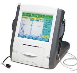
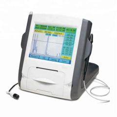
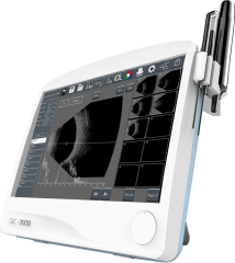
SW-2100 Ophthalmic Ultrasound AB Scanner


 SW-2100 user manual.pdf
SW-2100 software.pdf
SW-2100 user manual.pdf
SW-2100 software.pdf
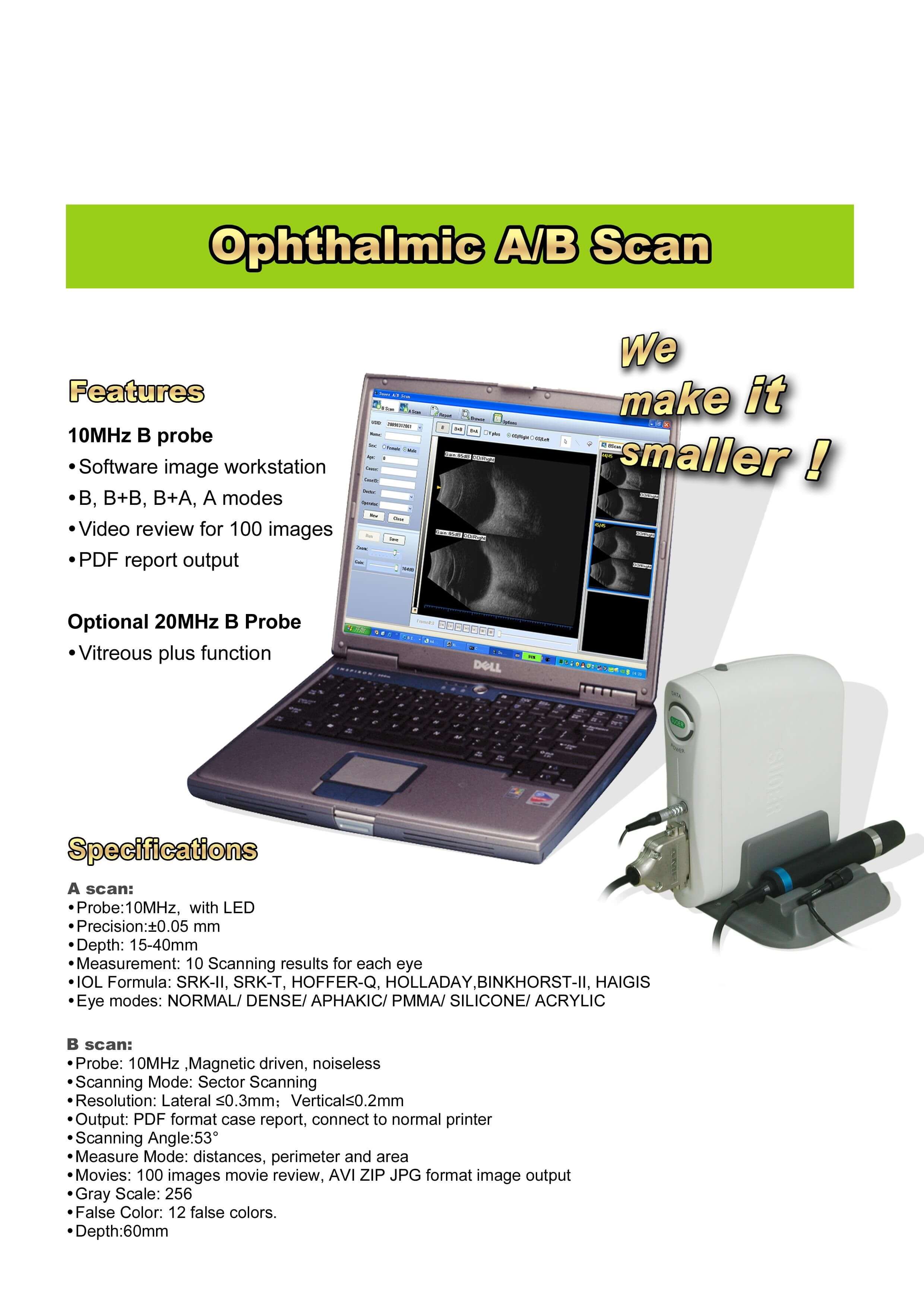
detail:
1.Software image workstation
2.B, B+B, B+A, A modes
3.Video review for 100 images
4.PDF report output
5.vitreous plus function
|
|
|
| A scan |
1.Probe: 10MHz frequencies, with LED |
| B scan |
1.Probe: 10MHz/20MHz (optional), Magnetic driven, noiseless |
| Others |
1.Display Mode :B, B+B, B+A, A |
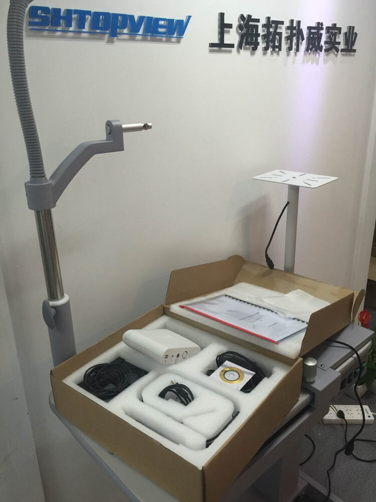


Shanghai Top View Industrial Co.,Ltd
Copyright © Shanghai Top View Industrial Co.,Ltd Technical by singoo
Privacy Policy