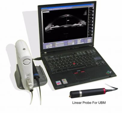
SW-3200S Full Scale Portable Ultrasound Bio-microscope
  • SW-3200.pdf SW-3200.pdf
  • Software Manual for UBM(081212).pdf Software Manual for UBM(081212).pdf
  • SW-3200 Full Scale Portable Ultrasound Bio-microscope

SW-3200 Full Scale Portable Ultrasound Bio-microscope free shipping

SUOER Full Scale UBM SW-3200s


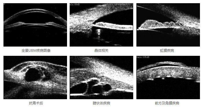
Linear scan range of up to 16mm; SW-3200S is small and easy for general examination and mobile examination. Used to check the anterior segment, structure display and lesions diagnosis, including cornea, chamber angle, ciliary body, iris, anterior chamber etc., double-angle display and measurement. Has been widely used in glaucoma, eye injury and ciliary body diseases.


Frequency: 50MHz
Scaning Mode: Wide range linear scanning mode, undistorted, Sulcus-to-Sulcus.
Scanning Range: 16mm*9mm; 10mm*6.5mm
Image Resolution(full screen):50um
Scaning Lines: 1024 lines, 15 um between each,high density & high resolution
Geometry Distortion: Both Vertical & Lateral rate<3%,no compensation, undistorted image
Probe: Multi-Dimension, precise locating articulated arm
Display Mode :UBM/UBM+A.
Magnify: Special independent ultrasound magnify system,better image in Anterior Segment.
Eye Fixation: Unique Eye position and LED system
Working Platform: Windows XP/VISTA/WINDOWS7
Image Process: Length, angle measurements
Output: Windows browser view, examining report can be created

Introduction and Clinic application
Introduction and Clinic application Introduction

SW-3200A Ophthalmic UBM is a professional ophthalmic ultrasound
image device. It is composed of main unit, 50MHz fan scan probe, image
workstation and so on.
This device is for ultrasound diagnose in hospital or clinic. Its main
application is detecting the anterior part of eye structure and disease, including
cornea, chamber angle, ciliary body, iris, anterior chamber and so on.
This device should be operated by trained doctor.

Clinic application of UBM

1 Anterior chamber angle detecting and form observing
2 Measure the depth of anterior and posterior chamber.
3 Detecting the thickness of iris, the form of tissue around iris and attach
position.
4 Get to know the form, position of lens and its relation with tissues around
5 Detecting the form and location of ciliary body
6 Diagnose whether the ciliary body or choroidea is off its place, and know
the degree of dropsy.
7 After the artificial lens is planted, its location can be observed by UBM.
8 For the eye injury, UBM could show the precise location of the injury on the
cornea and sclera, whether the chamber angle is set back , whether iris is
broken from the root or the lens is out of place. Also the UBM helps to
show the size and position of the eyewinker in the anterior eye.
9 UBM is helpful to the diagnosing and instruction therapy of coloring
glaucoma.
10 In case of angle closure glaucoma, it helps to know the theory of anterior
angle closure after the darkroom experiment.
11 For cornea opacity patients, UBM could detect the structure of cornea,

pupil and lens, which normal optical instrument could not detect. So the
UBM could perform normal examination before cornea plant operation

12 Valuate the form and function after the Glaucoma Filtration Surgery.
13 The diagnoses and identification of anterior segment cyst and solid tumor.

Contraindication of UBM Examination The following cases are forbidden to UBM examination:

1 Infective eye disease, such as Keratitis and so on.
2 Open eye damage, such as avulsion or perforation of cornea
3 Children under 5 years old that could not cooperate with the examination.

Environment Requirement    Characteristics Work Environment 1 Ambient temperature:+5°C ~ +40°C

2 Relative moisture:≤ 80%
3 Power Supply:AC 90~230V,50×(1±2%)Hz
4 Keep away from strong magnetic or electronic field

Transport and Storage

1 Ambient temperature:-40°C ~ +55°C
2 Relative moisture:≤ 95%;
3 Atmosphere pressure:500hPa~1060hPa

Running

1 Ambient temperature:+5°C ~ +40°C
2 Relative moisture:≤ 80%
3 Power Supply:AC 90~230V,50×(1±2%)Hz
4 Atmosphere pressure:860hPa~1060hPa
5 Warm-up time:5min

Basic Parameters

1 Probe:50MHz fan scanning UBM probe;
2 Vertical precision: 50µm;
3 Lateral precision: 50µm;
4 Scanning mode: fan scan, cornea protection, safty alarm;
5 Gain: 20-105dB;
6 SNR: 100dB;
7 Gray scale: 256 shades;
8 Display resolution: 0.01mm;
9 Micro mode 5mm×5mm and Full scale 12mm×10mm;
10 Geometry location precision: Vertical≤5%,Lateral≤10%;
11 Power supply: AC 90~230V,50×(1±2%)Hz;
12 Power : 10W;
13 Workstation OS: Windows XP,with USB2.0 interface,EMS
memory 512 MB;
14 Image store ability: 80G for image store in hard disk, and the store
ability could be expanded by CD burning.

Software Function

1 Long-time protection for probe: When the probe has been
continously working for 10 minutes, it will auto freeze and stop
scanning.
2 High Gain, High SNR, Wide dynamic range;
3 When the probe is blocked or too close to cornea, it will give out
alarm sound ,freeze automatically and stop scanning.
4 The functions are designed according to the procedure of
ophthalmic examination.The operation is simply and practical, fit for
doctor’s habbits.

5 Image display: Black&White, all kinds of false color.
6 Abundance of measure and after process software, recording 4
groups of distance, 2 groups of angles, perimeter and area.
7 Image output:all kinds of format including avi,zip and so on;insert
Windows browser whenever you like.
8 Real time image acquisition, auto record 100 frames in 10 seconds
before freezing. Images could be reviewed one-by-one or
continuously, convenient for doctors to have dynamic review.
9 Case output: provide PDF format case report with law validity,
while auto backup at the same time for future check up. The case
reports can be archived, searched or counted.
10 More than 20 kinds of eye location signals help to precisely locate
the focus.
11 Detailed professional dictionary makes the operation easier.

Safety

Accord with the requirement for GB9706.1-1995 Class I Type B
devices and GB9706.9


Our Services

Dear customer please leave your E-mail Address ,we `ll send you detailed user manual ,in order to let you know this instrument well

If you have any questions about our project, please feel free to contact us. We are happy to answer all your queries. E-mail will be answered, except weekends and holidays time. Since the time difference between shanghai and the location of your area, sometimes the answer will be delayed. Please be patient, We will respond you within 24 - 48hr(During holidays and bank holidays are un exception). and will give you a satisfied answer. if no response for you within 48 hours, please check the spam in your mail box. thank you


Product Catalog